The Complete Live-Cell Imaging Workflow

Quantitative live-cell analysis requires maintaining a stable physiological environment to preserve data fidelity. Protect cell viability while minimizing background fluorescence with a structured strategic workflow.

The 5-Step Roadmap

01 | PLAN

Design the experiment with careful consideration of spectral compatibility and consider longer-wavelength reagents to lower required excitation power.

02 | CULTURE

Maintain cells in optimum physiological conditions to prevent growth artifacts and metabolic perturbations.

03 | LABEL

Target specific cellular structures and functions with high-selectivity, low-toxicity probes, strictly avoiding excessive labeling concentrations.

04 | OPTIMIZE

Minimize extracellular background fluorescence and utilize specialized live-cell antifade reagents to maintain signal photostability.

05 | IMAGE

Capture kinetic discoveries with maximum clarity using imaging systems that offer precise control over light sources and exposure parameters.

Fluorescence Imaging of Live Cells
Solving the Technical Challenge

Live-cell fluorescence imaging enables the real-time observation of dynamic cellular processes. In contrast to fixed-cell assays, extended time-lapse imaging requires precise optimization of illumination parameters to prevent phototoxicity. High intensity excitation light generates reactive oxygen species (ROS), leading to metabolic shifts, membrane perturbations, and premature cell death.

Simultaneously, standard cell culture media components, specifically riboflavin and phenol red, produce significant background autofluorescence under standard excitation wavelengths. This optical background reduces the signal-to-noise ratio (SNR) and masks low-intensity biological signals. Eliminating these artifacts requires a structured optimization of imaging media, consumables, and fluorescent reagents to preserve cellular homeostasis during long-term experiments.

For a detailed methodology on mitigating these imaging artifacts, review the comprehensive guide below.

Step 1: Plan

Panel Configuration

Configure multiplexed panels using fluorophores with maximum spectral separation, and strictly optimize probe concentrations to avoid nonspecific staining, elevated background noise, and accelerated cytotoxicity caused by over-labeling. Additionally, carefully align your time-course duration and capture frequency with cellular tolerance to minimize cumulative light exposure.

Interactive Panel Design Tools

Evaluate channel compatibility, map absorption and emission spectra, and select optimized live-cell targets before initiating live-cell assays.

Step 2: Culture

Physiological Continuity & Environmental Stability

Maintaining absolute physiological homeostasis during multi-day kinetic imaging is critical to ensure that observed cellular behavior reflects native biology rather than environmental or optical artifacts. The choice of culture media, specialized live-cell cultureware, and precise environmental control directly dictates long-term cell morphology, growth rates, and metabolic health.

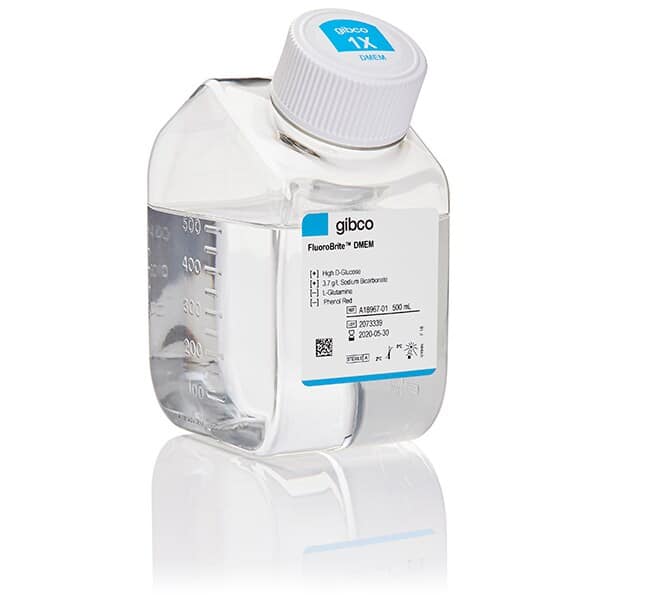
FluoroBrite™ DMEM Media

Standard formulations containing phenol red and riboflavin generate high background autofluorescence under illumination. FluoroBrite™ DMEM eliminates this spectral noise to reduce background by up to 90%, enabling high-contrast imaging at reduced excitation intensities to protect cell viability.
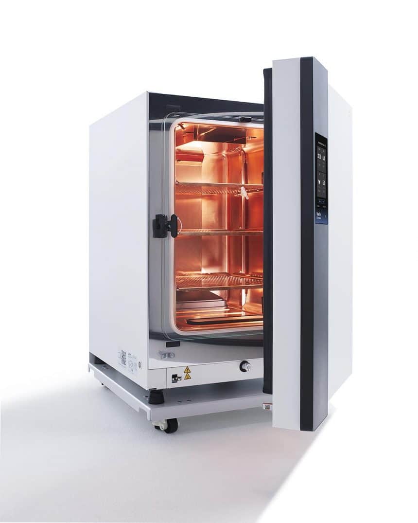
Vios™ iDx Precision Incubation

Long-term kinetics for incubator-based imaging systems (such as the Sartorius® Incucyte®) require pristine atmospheric stability to prevent growth artifacts. Vios™ iDx incubators deliver rapid CO₂ recovery and extreme thermal uniformity to shield sensitive cells from ambient fluctuations.
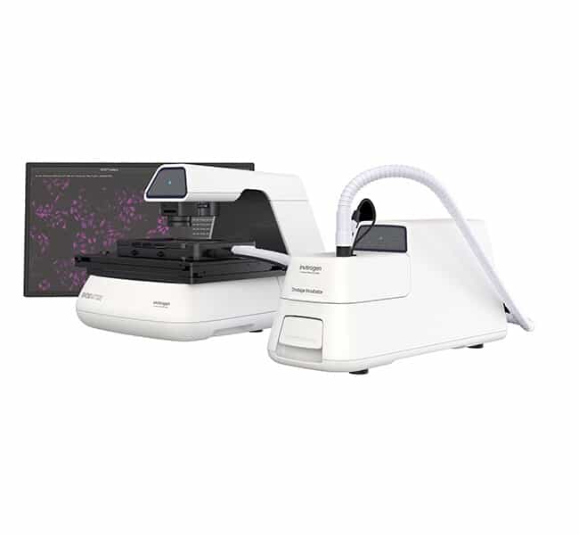
EVOS™ & CellInsight™ Onstage Incubators

For stage-top imaging workflows, environmental parameters must be tightly regulated directly on the microscope stage. EVOS™ and CellInsight™ Onstage Incubators function as precise environmental chambers, enabling strict control of temperature, humidity, and gases during intensive time-lapse acquisition.
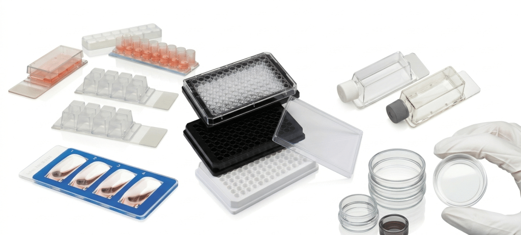
Nunc™ Validated Imaging Consumables

Standard tissue culture vessels can introduce optical distortion and background autofluorescence. Nunc™ cell culture imaging consumables feature high-quality glass or optical polymer bases to ensure optimal refractive indices, formal support for automated high-content screening, and seamless compatibility with live-cell imaging platforms.

Step 3: Label

Reagent Selection Guide

Live-cell imaging reagents include both targeted fluorescent proteins and small membrane-permeant fluorescent dyes. To enable live-cell imaging experiments, many reagents are designed for time-lapse over several hours or days, while others are optimal for end-point assays in which cells are imaged and analyzed immediately after staining. Staining concentration, incubation time, and the appropriate time window and imaging interval/frequency should be determined empirically to minimize cytotoxicity and preserve cellular function.

The reagents found within the live-cell imaging guide below are compatible with automated high-content and incubator-based fluorescence imaging systems, such as the EVOS™ M7000 or EVOS™ M5000 Imaging Systems with EVOS™ Onstage Incubator, CellInsight™ HCS Platforms, PerkinElmer’s MuviCyte™ live-cell imaging system*, Leica’s Thunder and Mica*, and Sartorius’ Incucyte® Live-Cell Analysis System™*.

Biological Target / ApplicationInvitrogen™ SolutionPrimary Filter / Cube MatchImaging Timeframe & Duration
Viability (Live Cell Indicators)Calcein AM / Calcein Blue, AMGFP / DAPIShort-term (minutes to hours)
Viability (Dead Cell Indicators)SYTOX™ Green, SYTOX™ Orange, SYTOX™ Deep Red / NucGreen™ Dead 488 ReadyProbes™ / YOYO™-1 / YOYO™-3 / Image-iT™ DEAD Green Viability StainGFP / Orange / Red / NIRShort- to long-term (24+ hours)
Viability / Cytotoxicity KitsLIVE/DEAD™ Viability/Cytotoxicity Assay Kit (Green/Deep Red)GFP / NIRShort-term (minutes to hours)
Apoptosis (Caspase-3/7)CellEvent™ Caspase-3/7 Green Detection Reagent / CellEvent™ Caspase-3/7 Red Detection Reagent / Annexin V ConjugatesGFP / RFPLong-term (overnight to 48–72 hours)
AutophagyPremo™ Autophagy Sensors (p62 and LC3B) GFP / Premo™ Autophagy Sensors (p62 and LC3B) RFPGFP / RFPLong-term (overnight to 48 hours)
Cell CycleCellLight™ FUCCI Cell Cycle IndicatorGFP / RFPLong-term
Biological Target / ApplicationInvitrogen™ SolutionPrimary Filter / Cube MatchImaging Timeframe & Duration
Reactive Oxygen Species (ROS)CellROX™ Green Reagent / CellROX™ Orange Reagent / CellROX™ Deep Red ReagentGFP / Orange / NIRLong-term (24 hours)
General Oxidative StressH2DCFDA dyesGFPShort-term (~2 hours)
Mitochondrial SuperoxideMitoSOX™ Green Superoxide IndicatorGFPEnd-point
Thiol Status EvaluationThiolTracker™ VioletDAPI / VioletEnd-point
Hypoxia MonitoringImage-iT™ Green Hypoxia ReagentGFPEnd-point
Calcium SignalingFluo-4 Calcium Imaging Kit / Rhod-3 Calcium Imaging KitGFP / RFPShort-term (minutes to hours)
Biological Target / ApplicationInvitrogen™ SolutionPrimary Filter / Cube MatchImaging Timeframe & Duration
Antibody Binding AssaysZenon™ Alexa Fluor™ Plus 488 / Zenon™ Alexa Fluor™ Plus 594 / Zenon™ Alexa Fluor™ Plus 647GFP / RFP / NIRShort-term
Antibody InternalizationpHrodo™ Green iFL / pHrodo™ Red iFL / pHrodo™ Deep Red Antibody Labeling Reagents / LysoLight™ Green & LysoLight™ Deep Red Labeling KitsGFP / RFP / NIRLong-term (hours to overnight / ~72 hours)
Phagocytosis DynamicspHrodo™ Green, Red, and Deep Red BioParticles™ ConjugatesGFP / RFP / NIRShort- to long-term (minutes to hours or longer)
Endosomes / EndocytosispHrodo™ and Alexa Fluor™ dextransGFP / RFP / NIRShort- to long-term (minutes to hours; overnight or longer)
Ligand InternalizationAlexa Fluor™, BODIPY™, and pHrodo™ LDL / EGF / Transferrin conjugatesGFP / RFP / NIRShort- to long-term (minutes to overnight or longer)
Biological Target / ApplicationInvitrogen™ SolutionPrimary Filter / Cube MatchImaging Timeframe & Duration
Mitochondria StructureMitoTracker™ dyesGFP / Orange / RFP / NIRLong-term
Mitochondria FunctionTMRE / TMRMOrange / RFPShort-term (minutes to hours)
Cytoskeleton (Actin)CellMask™ Green, Orange, and Deep Red Actin Tracking StainsGFP / Orange / NIRShort- to long-term (24+ hours)
Cytoskeleton (Tubulin)Tubulin Tracker™ Green / Tubulin Tracker™ Deep RedGFP / NIRShort-term
Plasma MembraneCellMask™ Green, Orange, and Deep Red Plasma Membrane StainsGFP / Orange / NIRShort-term (10–90 minutes, before internalization)
Membranes (Plasma & Internal)Vybrant™ cell labeling solutionsGFP / Orange / RFP / NIRLong-term
Endoplasmic ReticulumER-Tracker™ Green / ER-Tracker™ RedGFP / RFPEnd-point
LysosomesLysoTracker™ and LysoSensor™ dyesGFP / RFP / NIRShort-term (minutes to hours)
Nucleus MorphologyHoechst 33342 / NucBlue™ Live / NucRed™ Live 647 / DRAQ5™ / SYTO™ 9 / SYTO™ 59 / HCS NuclearMask™ Blue, Red, and Deep Red StainsDAPI / GFP / RFP / NIREnd-point or Live
Cell Tracking / ProliferationCellTracker™ dyes / Vybrant™ CFDA SE Cell Tracer KitGFP / Orange / RFP / NIRShort- to long-term (72 hours / 3+ generations)
Protein-Based Target TrackingCellLight™ GFP BacMam 2.0 / CellLight™ RFP BacMam 2.0 Structure ReagentsGFP / RFPLong-term (overnight to days / 2–3 generations)

Live-Cell Imaging Resources

Download the comprehensive Molecular Probes™ Handbook and access practical imaging protocols and selection guides to optimize your fluorescent labeling configurations.

Step 4: Optimize

Signal-to-Background Ratio & Photostability

Signal-to-background ratio can be optimized by using specialized reagents designed to reduce extracellular fluorescence and increase fluorophore photostability during intensive time-lapse acquisition.

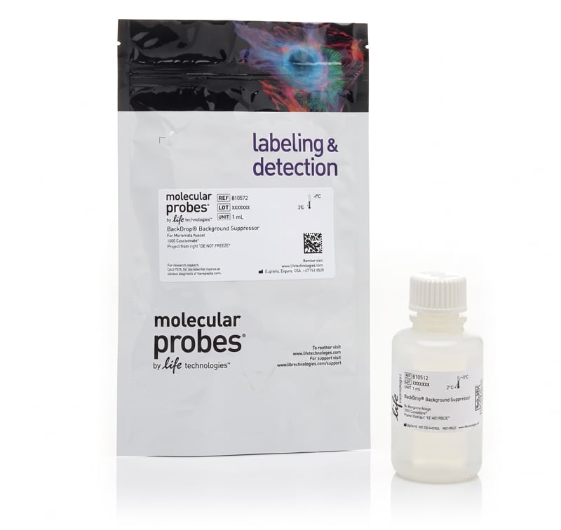
Invitrogen™ BackDrop™ Background Suppressor

Used when observing high background signal or weak fluorescence in the blue, green, or red channels. The addition of this live-cell compatible background suppressor reduces extracellular autofluorescence and eliminates the need for a wash step, enabling a high-contrast, no-wash protocol.

Invitrogen™ ProLong™ Live Antifade Reagent

Applied directly to samples to increase fluorophore photostability and decrease the cumulative effects of phototoxicity across a variety of sample types. Based on protection metrics under intensive illumination, it permits up to 100% more image captures. Following 120 exposures in standard time-lapse protocols, treated samples remain >20% brighter than untreated cells, extending data collection windows.

Step 5: Image

Precision Acquisition & Quantitative Analysis

Optimized live-cell data collection relies on imaging hardware engineered to maximize signal capture while minimizing light exposure. Preserving true biological responses over extended kinetic time-courses requires precise illumination timing and automated focal stability.

EVOS™ M5000 & M7000 Imaging Systems

Benchtop microscopes utilizing independent, adjustable EVOS™ LED Light Cubes (DAPI, GFP, RFP, Red, and Cy5 configurations) to deliver targeted, brief illumination strictly during active acquisition windows. This avoids continuous sample exposure and significantly protects cell viability during long-term time-lapse experiments when integrated with the EVOS™ Onstage Incubator.

CellInsight™ High-Content Screening (HCS) Platforms

Automated, high-throughput imaging systems combining high-resolution, artifact-free optics with rapid multi-channel scanning. These platforms enable researchers to track complex structural dynamics, metabolic functions, and cellular health across entire multi-well configurations with absolute reproducibility.

Workflow Consultation
Navigating the complexities of live-cell imaging requires a coordinated approach across every step.

Our application specialists are available to consult on environmental optimization, instrumentation, and reagent selection to ensure your research goals are achieved with precision.