

EVOS M3000 Imaging System
Compact, connected, and easily upgradable
The EVOS M3000 Imaging System is a fully integrated digital inverted benchtop microscope for two-color fluorescence, transmitted light, phase contrast, and color imaging. It is excellent for routine cell/tissue culture imaging applications. This system combines an easy user interface with powerful machine learning capabilities that allow users to quickly verify cell appearance, quality, and growth in seconds.
The EVOS M3000 offers you these important advantages:
- Easy installation—no maintenance, assembly, alignment, or calibration
- Automated real-time confluency analysis—includes pre-trained machine learning algorithm that can report cell confluency in the field of view at any time. As vessel is moved, confluency value automatically updates (in under a second on average).
- User-defined two-color fluorescence—with addition of EVOS light cubes, two fluorescence channels can be acquired
- Space-saving design—at 12 x 19 x 13 in. (W x D x H), system fits easily on lab bench or in cell culture hood
- Network-capable—can be connected to local wireless network for seamless data transfer to OneDrive™ or Thermo Fisher Connect accounts. Alternatively, system offers on-board storage or export to USB.
- Flexibility—choose from wide selection of EVOS-compatible objectives (1.25–60X) to best fit application and image quality needs.
GO TO SHOP – Cell Analysis Products
EVOS real-time confluency tool
EVOS real-time confluency tool can typically measure confluency in less than 1 second without requiring any image capture, making it excellent for eliminating user bias in routine cell culture.
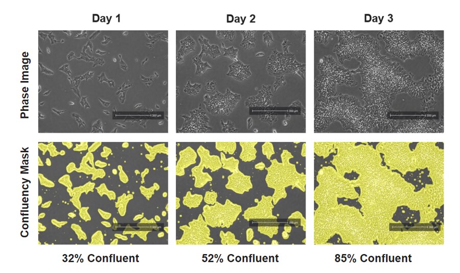
Induced pluripotent stem cells imaged over time with the EVOS M3000 instrument. Human fibroblast-derived induced pluripotent stem cells were cultured on a vitronectin-coated 6-well plate in Gibco Essential 8 Flex Medium for 3 days. Cells were imaged with the EVOS M3000 instrument under phase microscopy with and without the automatically generated confluency mask and measurement.
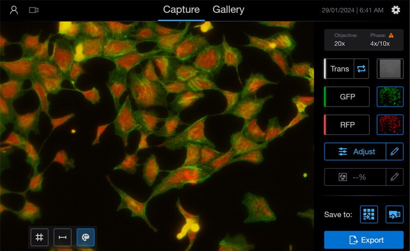
Powerful, intuitive software
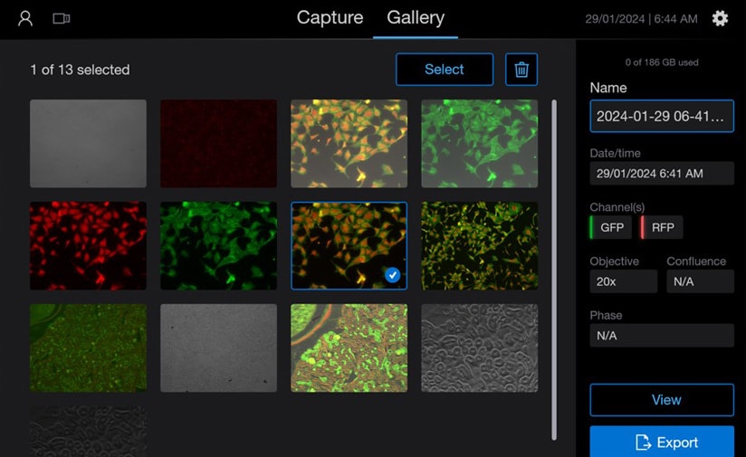
Users can easily share files from the instrument via the Thermo Fisher Connect cloud ecosystem, as well as MS OneDrive, making it an exceptional solution for research teams.
As with every EVOS instrument, overlay scale bars, gridlines, or adjust brightness, saturation, and gamma correction prior to imaging.
Following acquisition, scroll through all images and video in a gallery view to tag those of interest or easily delete any you don’t want.
| Manufacturer |
|---|


